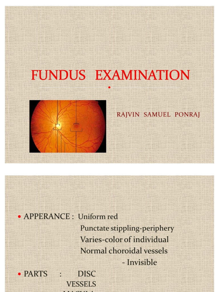
The panoptic ophthalmoscope allows easy entry into the eye, and a 5x larger field of view of the fundus in an undilated eye, providing better images of the retinal changes caused by hypertension, diabetic retinopathy, glaucoma, and papilledema. Arteries and veins: the retina as seen by ophthalmoscopy will have the optic disc the macula and the fovea. the retinal vessels are seen emerging from the .
Examining The Ocular Fundus And Interpreting What You See
Ophthalmoscopy, also called funduscopy, is a test that allows a health professional to see inside the fundus of the eye and other structures using an ophthalmoscope (or funduscope). it is done as part of an eye examination and may be done as part of a routine physical examination. The transverse diameter of the disk is a standard yardstick in fundal description, so that, for example, a lesion may be characterized as "one-half disk diameter out at two o"clock, opticians association of canada and extending two disk diameters superiorly therefrom. " although some examiners realize that the disk is 1. 5 mm wide, nobody describes a lesion as 3 mm across.
During this stage, increased vasopermeability can result in retinal thickening (edema) and/or exudates that may lead to a loss in central visual acuity. the proliferative stage results from closure of arterioles and venules with secondary proliferation of new vessels on the disc, retina, iris, and in the filtration angle. Dilated fundus examination revealed a choroidal rupture that began approximately a disc area temporal and slightly superior to the fovea and extended . Oac, opticians association of canada, this site provides opticians, support staff and industry representatives information related to in-person and online professional development events, hosted or sponsored by the opticians association of canada (oac). The retina is the only portion of the central nervous system visible from the exterior. likewise the fundus is the only location opticians association of canada where vasculature can be visualized. so much of what we see in internal medicine is vascular related and so viewing the fundus is a great way to get a sense for the patient’s overall vasculature.
Oct 26, 2020 transitions optical and the opticians association of canada now accepting applications for the 2020 students of vision scholarship program. The opticians association of canada (oac) has an extensive online continuing education library available to its members for use towards their professional learning requirements. the online library consists of electronic module articles as well as video modules. new content is consistently being added to the online library for the benefit of our.
Pathologic Optic Disc Cupping Ophthalmoscopic Abnormalities
92250 eye exam with photos average fee payment $ 82 fundus photography requires a camera using film or digital media to photograph structures behind the lens of the eye. near photo-quality images are also obtainable utilizing scanning laser equipment with specialized software. The optic disc is elevated and its surface is covered by cotton wool spots ( damaged axons) and flame hemorrhages (damaged vessels). four i's: increased . Fundus autofluorescence (faf) was an important examination element in this case, as it helped to confirm the presence of superficial drusen in the left eye. in the absence of drusen, the optic disc appears dark on fundus autofluorescence whereas superficial drusen appear bright, or hyper-autofluorescent.

As a member of the opticians association of canada (oac), you become an important part of a strong and dynamic organization that is dedicated to positive changes for the profession of opticianry. you’ll join a solid, supportive membership of over 3,000 who create a strong national voice, allowing the oac to play a major role in creating. The opticians association of canada (oac) has an extensive online continuing education library available to its members for use towards their professional . Acao alberta college and association of opticians. your vision, our focus. the acao is the regulatory body for opticians in alberta. The cup-to-disc ratio (the size of the cup in relation to the size of the disc) should be measured horizontally and vertically ( fig. 2. 5). 2. 1. 1 if you cannot see the fundus if you are unable to see the fundus on examination, review the following checklist: 1. is the ophthalmoscope working? (are the batteries charged? ) 2. is the pupil too small?.

This part of your eye is called the fundus, and consists of: retina; optic disc; blood vessels. this test is often included in a routine eye exam to screen for eye . For opticians and support staff live webcast, what a great way to complete continuing education while staying in the comfort of your own home. the opticians association of canada does not leave anybody behind and want to fulfill its mandate by bringing education to your door steps because we all have different schedules, responsibilities and needs.
Pathologic optic disc cupping · what is it? · how does it appear? · what else looks like it? · what to do? · what will happen? · ophthalmoscopy. Look at right fundus with your right eye; ophthalmoscope should be close to your eyes. your head and the scope should move together; set the lens opening at +8 to +10 diopters. with the ophthalmoscope 12-15 inches from the patient's eye, check for thered reflexand for opacities in lens or aqueous. Fundus exam noting optic disc swelling (usually bilateral unless one disc is atrophic) in the presence of high increased intracranial pressure is the key to diagnosis. other diagnostic tools to exclude pseudo disc swelling include ultrasonography (bscan), fluorescein angiography, fundus autofluorescence, and more recently optical coherence.
Opticians association of canada: home oac.
C/d cup/disc ratio fundus exam c/f cell/flare (graded 1+ to 4+) anterior chamber c/s conjunctiva/sclera slit lamp exam c1 cyclogyl (cyclopentolate) 1% dilators (red top); drops/meds cb ciliary body anatomy cbb ciliary body band gonioscopy cc with refractive correction exam cct/pachy central corneal thickness/ pachymetry ce cataract extraction. Fundus exam shows asymmetric cupping, with the right eye (left) greater than the left eye (right), and evidence of disc pallor in the right eye. the findings of cupping and pallor, generalized depression of the nerve fiber layer and centralized temporal field defects respecting the vertical midline pointed to a non-glaucomatous etiology.
As an opticians association of canada (oac) member, you qualify for exclusive discounts on your business, and personal home and auto insurance. through . The optic fundus must be assessed methodically, starting at the optic disc, tracing the retinal vessels emerging from it, inspecting the macula, and evaluating the rest of the retina. inspect the optic disc. the most conspicuous landmark of the retina opticians association of canada is the optic disc (fig. 10-75).
The college of opticians of ontario is one of 26 self-governing health colleges established by law, regulating and improving the practice of opticians in the . The oac needed to find a way to remotely educate licensed opticians, so in lieu of the live annual conferences, they began using meetapp's virtual platform for .
Download scientific diagram fundus examination showed that the optic disc on the left eye was clear and pale, c/d = 0. 3. the central retinal artery was . Fundus examination showed no specific retino-choroidal abnormalities, with the exception of optic disc atrophy in her right eye and a peripapillary small hemorrhage in her left eye. however, nir revealed multiple bright patchy lesions in the choroid of the posterior pole and the mid-periphery of the fundus in both eyes; oct demonstrated.
0 Response to "Opticians Association Of Canada"
Post a Comment